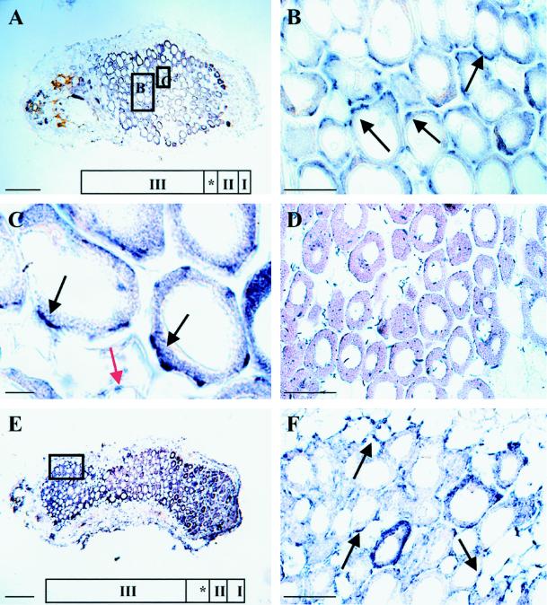
Figure 2.
Localization of the NADH-GOGAT protein in longitudinal sections of 19-d-old (A–C) and 33-d-old (E and F) root nodules. Nodule ultrastructure was classified based on the nomenclature of Vasse et al. (1990): the meristem (zone I), the invasion zone (zone II), the interzone (*), and the N2-fixing zone (zone III). Sections A through E were probed with affinity-purified NADH-GOGAT antibodies and with alkaline-phosphatase-conjugated secondary antibodies. The signal, which reflects the localization of the NADH-GOGAT protein, is seen as blue spots. The black arrows point toward infected cells, and the red arrow points toward uninfected cells. Longitudinal section of a 19-d-old alfalfa root nodule is shown in A. Enlargement of the boxed regions in A shown in B and C includes part of the N2-fixing zone (zone III) with infected and uninfected cells. D shows control, affinity-purified NADH-GOGAT antibodies that were incubated together with the partially purified NADH-GOGAT polypeptide on a longitudinal section through a 19-d-old root nodule. E shows a longitudinal section through a 33-d-old root nodule, and F shows an enlargement of the boxed region in E that is part of the proximal region of the nodule. Bars in A and E = 270 μm; bars in B, D, and F = 70 μm; and bar in C = 15 μm.

