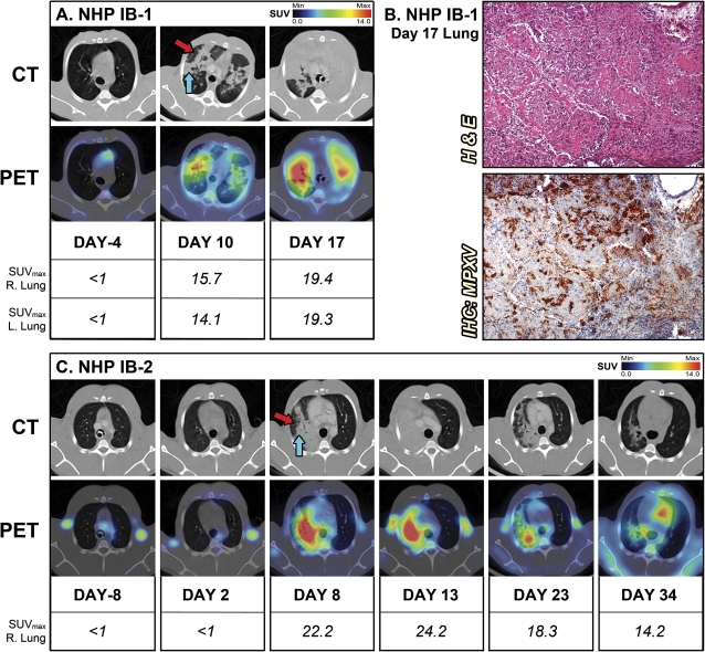
Figure 1.
PET/CT) imaging of lung inflammation. A, For the nonhuman primate (NHP) IB-1 that succumbed to infection, sequential axial lung sections were imaged on day –4 and days 10 and 17 after infection. B, Histopathology and monkeypox virus (MPXV) antigen reactivity of lung on day 17. C, Sequential axial lung sections were imaged in surviving NHP IB-2 on day –8 and days 2, 8, 13, 23, and 34 after infection. (Red arrow) Hazy attenuation. (Blue arrow) Consolidated area. Maximum standard uptake values (SUVmax) for fluorine-18 fluorodeoxyglucose (18F-FDG) in right and left sections of lung were determined on scanning days by PET. The PET/CT slice that was chosen for each image corresponds to the maximum SUV value determined in the volume of interest (VOI) of the lung. Abbreviations: H & E, hematoxylin and eosin; IB, intrabronchial; IHC, immunohistochemistry.

