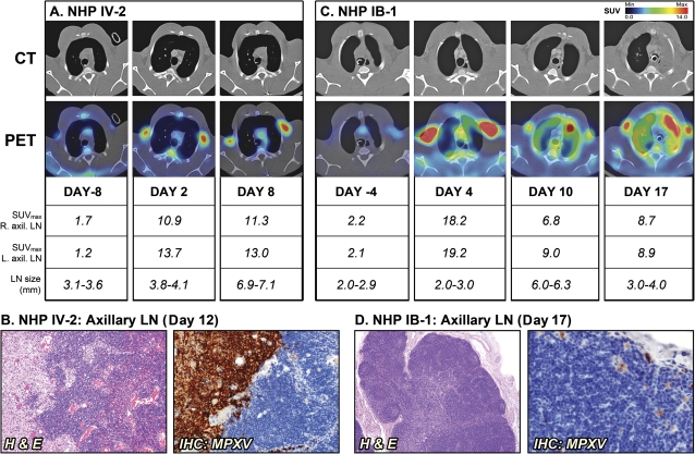
Figure 2.
Sequential positron emission tomography/computed tomography (PET/CT) imaging of axillary lymph nodes (LNs) in nonhuman primates (NHPs) that succumbed to disease. A, NHP IV-2 was scanned on day –8 and days 2 and 8 after infection (d12, day of death). B, Histopathology of NHP IV-2 on day 12. C, NHP IB-1 was scanned on days –4 and days 10 and 17 after infection (day 17, day of death). D, Histopathology of NHP IB-1 on day 17. Fluorine-18 fluorodeoxyglucose (18F-FDG) maximum standard uptake values (SUVmax) and short axis measurements of axillary LNs (LN size) were determined on scan days by PET and CT imaging, respectively. The PET/CT slice that was chosen corresponds to the maximum SUV value of the LNs. Abbreviations: H & E, hematoxylin and eosin; IB, intrabronchial; IHC, immunohistochemistry; IV, intravenous; MPXV, monkeypox virus.

