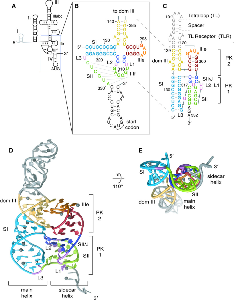
Figure 1. Structure of the HCV IRES pseudoknot domain.
(A) Secondary structure cartoon of the HCV IRES with names of major domains indicated. Dom I is shown in light grey as it is not considered part of the IRES and is not present in the reporter constructs used here. (B) Pseudoknot-domain secondary structure, as conventionally drawn. Loops and stems are indicated by L and S, respectively. (C) Crystallization construct of pseudoknot domain, with secondary structure redrawn according to the crystal structure. Nucleotides from the IRES are numbered as in the IRES and the crystallization-module nucleotides are numbered 1–21. Crystal structure shown (D) head-on and (E) at a 110° angle, looking down the sidecar helix. Ni2+ ions are shown as grey spheres. Secondary structural elements are colored consistently in b–e (dom III, yellow; SI, light blue; IIIe, red; SII/J, deep blue; and SII, green) and the crystallization module (tetraloop, spacer, tetraloop receptor) is shown in grey. The ‘main’ and ‘sidecar’ helices consisting of dom III + SI and IIIe+SII/J+SII, respectively, are labeled. Pseudoknot (PK) 1 is composed of loop IIIf and downstream sequence and PK2 is formed between the IIIe tetraloop and the main helix of dom III. See also Figure S1.

