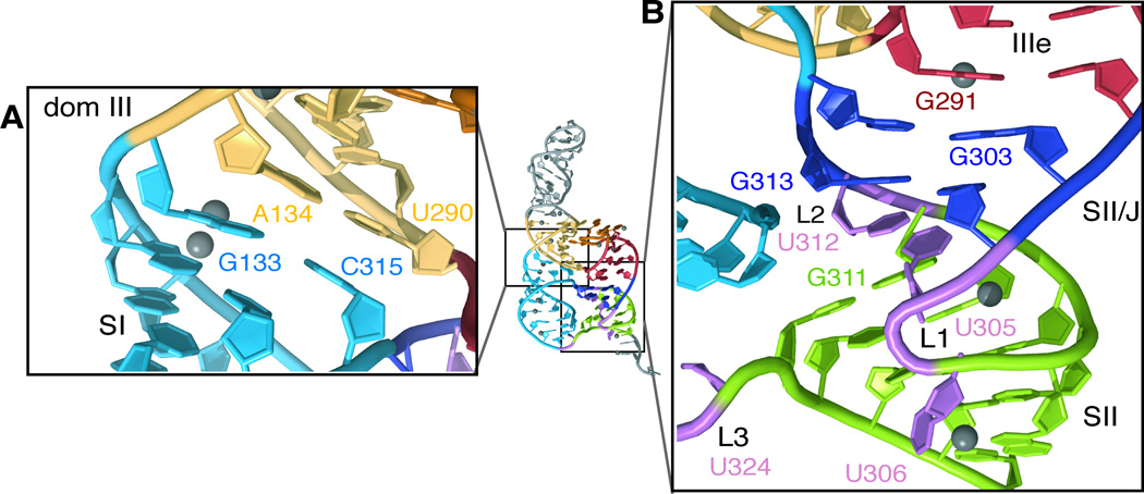
Figure 2. Structure of the IIIef four-way junction.
(A) Close-up of coaxial stacking between the main stem of dom III (yellow) and SI (blue). (B) Close-up of complex coaxial stacking within the sidecar helix, between IIIe (red), SII/J (deep blue) and SII (green). Note that the base of U324 (L3) is omitted from the crystallographic model, as no density was observed for this nucleotide apart from the sugar-phosphate backbone. Ni2+ ions are shown as grey spheres.

