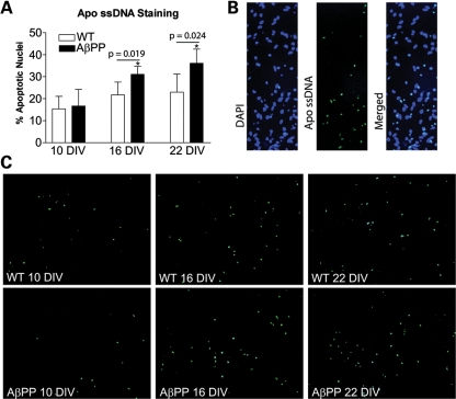
Figure 4.
Primary neurons from AβPP mice undergo premature apoptotic cell death compared with WT neurons. (A) Cells stained with the Apo-ssDNA antibody for apoptosis were counted and normalized to DAPI-stained nuclei in order to assess the percentage of apoptotic nuclei (n = 5 cultures from independent pups per group). (B) Apo-ssDNA-stained cells colocalized with pyknotic nuclei from DAPI stain. (C) Representative images of Apo-ssDNA-stained cultures from WT and AβPP-derived cultures harvested at 10, 16 and 22 DIV.

