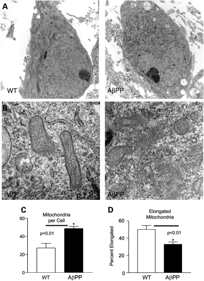
Figure 7.
Transmission electron microscopy analysis of cell body mitochondria shows increased numbers of rounded mitochondria in AβPP neurons. (A) Representative images of cell bodies are shown from AβPP-derived and WT primary neuronal cultures at 16 DIV. (B) Close examination reveals that many mitochondria in AβPP neurons lack clear membrane structure and appear fragmented. (C) Mitochondria per cell body were counted. n ≥ 10 images per group. (D) Elongated mitochondria were counted and normalized to total mitochondria to calculate the percent of elongated mitochondria. n ≥ 10 images per group.

