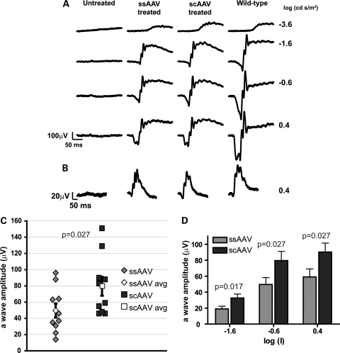
Figure 2.
Light-dependent photoreceptor responses from Aipl1−/− mice following early AAV treatment. One eye of Aipl1−/− mice was injected at P2 with the indicated AAV and ERGs were recorded at P30. ERG responses from (A) dark- and (B) light-adapted conditions were measured at the indicated log (cd · s/m2) flash intensities. Traces from a contralateral untreated eye and an age-matched wild-type mouse are shown for comparison. (C) Dark-adapted photoresponses from Aipl1−/− treated mice at the flash intensity of −0.6 (log cd s/m2). Each data point represents the measured a-wave amplitude of an individual treated mouse. The group average of scAAV-treated mice was significantly higher than the group average of ssAAV-treated mice (P ≤ 0.027). (D) Comparison of mean rod photoreceptor response (a-wave amplitude) between ssAAV and scAAV treatments over increasing flash intensities. Average a-wave amplitudes from the scAAV-treated mice were significantly greater than the ssAAV-treated mice at all intensities (P ≤ 0.027). Error bars, ±SEM.

