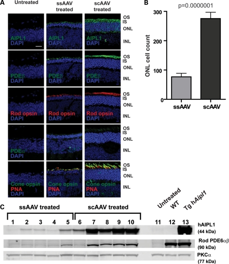
Figure 7.
Photoreceptor morphology of Aipl1−/− retina following late administration of AAV-mediated gene replacement. Aipl1−/− mice were treated at P10 and retinas were collected at P35 for immunocytochemistry. (A) Confocal images of retinal sections stained with indicated antibodies. Peanut agglutinin (PNA) (red) is used as a cone marker. Cell nuclei are stained with DAPI (blue). Images were taken at ×63 magnification. Scale bar, 20 µm. OS, outer segment; IS, inner segment; ONL, outer nuclear layer; INL, inner nuclear layer. (B) Quantification of photoreceptor cell nuclei showed significantly greater nuclei in scAAV- compared with ssAAV-treated retina (P ≤ 0.0000001). Error bars, ±SEM. (C) Western blots of ssAAV (lanes 1–5) and scAAV (lanes 6–10) treated retina. Untreated Aipl1−/− (lane 11), wild-type (lane 12) and transgenic hAipl1 (lane 13) retina serve as controls. PKCα expressed in bipolar cells, is a loading control.

