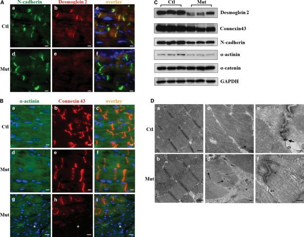
Figure 5.
Analyses of cell–cell junctions in 2-month-old Jup mutant mice. (A) Desmoglein 2 staining signal was lost at the Jup mutant intercalated discs (ICDs), whereas N-cadherin staining was similar between Jup mutant and control hearts. Bar, 10 μm. (B) Connexin 43 staining was normal in Jup mutant cardiomyocytes, but lost in the scar area (asterisk). Bars, 10 μm. (C) Western blot analyses of cell junction proteins. (D) TEM analyses reveal normal sarcomere structure in Jup mutant cardiomyocytes (b). Replacement of cardiomyocytes by collagen deposition (asterisk) in Jup mutant hearts (d). Black arrows point to the cardiomyocyte nuclei. (e) and (f) Normal adherens junction (asterisk) and gap junction (white arrows) at Jup mutant ICDs, whereas no desmosomes are identified in Jup mutant hearts, as opposed to typical desmosomes (black arrow) in control hearts. Bars (a and b), 500 nm; bars (c and d): 5 μm; bars (e and f): 250 nm.

