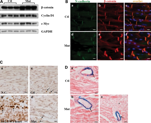
Figure 6.
Increased β-catenin at cell adhesion without affecting Wnt/β-catenin signaling in Jup mutant mice. (A) Western blot of total β-catenin and its signaling targets—Cyclin D1 and c-Myc. (B) Immunofluorescence staining of β-catenin in 18-day-old Jup mutant and control hearts. Note marked increase in expression of β-catenin in Jup mutant ICDs compared with that in control ICDs, no signal was detectable in nuclei or cytosols in either Jup mutant or control cardiomyocytes. Bars, 10 μm. (C) Immunostaining of Cyclin D1 in Jup mutant and control hearts (18 days old). (a) Serves as a negative control (without primary antibody incubation). (b) Positive Cyclin D1 staining (dark brown staining in nuclei indicated by arrows) is shown in embryonic day 12.5 (E12.5) WT cardiomyocytes. Positive stainings (arrows) are identified in some non-cardiomyocytes in both control (c) and Jup mutant (d) hearts. These non-cardiomyocytes are different from cardiomyocytes by presenting non-rod shape nuclei and localizing at the muscle bundle periphery. Note that no positive stainings were detectable in either control or Jup mutant cardiomyocytes. Bars, 20 μm. (D) Representative pictures of X-Gal staining of a 18-day-old Jupf/f/Fos-LacZ heart (a) and a 18-day-old Jupf/f/αMHC-Cre/Fos-LacZ heart (b and c). There is no signal in cardiomyocytes in either control or Jup mutant cardiomyocytes, nor is in scar tissues (asterisk) (c). In contrast, X-Gal staining signals (arrows) are apparent in vascular cells. Bars, 20 μm.

