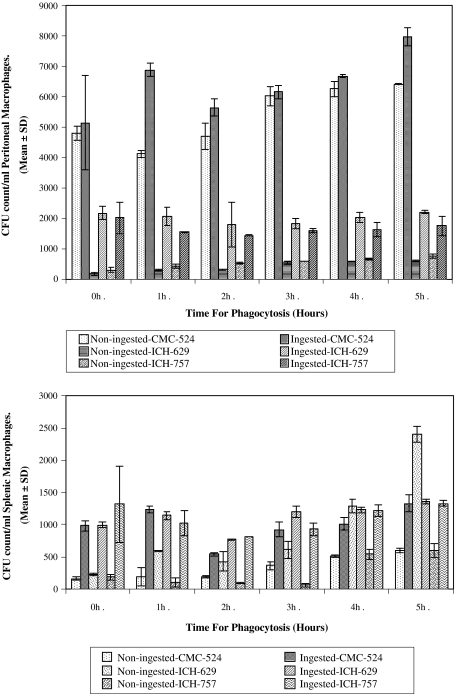
Fig. 2.
Phagocytosis of S. aureus by a murine peritoneal macrophages and b murine splenic macrophages. The intracellular killing of S. aureus by murine peritoneal macrophages after time dependent phagocytosis (up to 5 h) is shown in Fig. 2a. The results in this figure represent the gradual increased intracellular viability of S. aureus within murine peritoneal macrophages after 2 h of phagocytosis. The intracellular killing of S. aureus by murine splenic macrophages after time dependent phagocytosis (up to 5 h) is shown in Fig. 2b. The results in this figure represent the gradual increased intracellular viability of S. aureus within murine splenic macrophages after 2 h of phagocytosis

