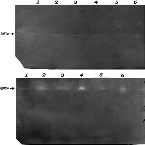
Fig. 5.
Detection of a SOD activity and b catalase activity in the acrylamide gels. Electrophoresis was performed on non-denaturing acrylamide gels using the Bio-Rad Mini system with tris/glycine buffer. Results in this figure represent the non- denaturing polyacrylamide gel electrophoresis (PAGE) analysis of SOD activities in the sonicated extracts of S. aureus (CMC-524, ICH-629 and ICH-757) in 12% native polyacrylamide gels. 1 CMC-524, 2 h lysate, 2 CMC-524, 4 h lysate, 3 ICH-629, 2 h lysate, 4 ICH-629, 4 h lysate, 5 ICH-757, 2 h lysate, 6 ICH-757, 4 h lysate. Electrophoresis was performed in a way as mentioned in methods. Regions corresponding to catalase activity were determined as yellow bands on a green background. Results in this figure represent the non- denaturing polyacrylamide gel electrophoresis (PAGE) analysis of catalase activities in the sonicated extracts of S. aureus (CMC-524, ICH-629 and ICH-757) in 8% native polyacrylamide gels. 1 CMC-524, 2 h lysate, 2 CMC-524, 4 h lysate, 3 ICH-629, 2 h lysate, 4 ICH-629, 4 h lysate, 5 ICH-757, 2 h lysate, 6 ICH-757, 4 h lysate

