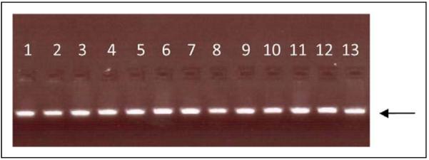
Figure 1.

Gel electrophoresis (1.5% agarose gel) of the PR-M localization sequence demonstrating isolation of the 48 base-pair PCR amplification product used for sequencing. The arrow indicates the gel position of 50 base pairs (15 ng). Thirteen samples are shown, with 1 sample per lane. PR-M indicates mitochondrial progesterone receptor; PCR, polymerase chain reaction.
