
Fig. 4. Interactions between Jagged1 and P-Erk1/2, P-Smad1/5/8, Wnt-β-catenin, and ephrinA-EphA pathways in the coronal suture.
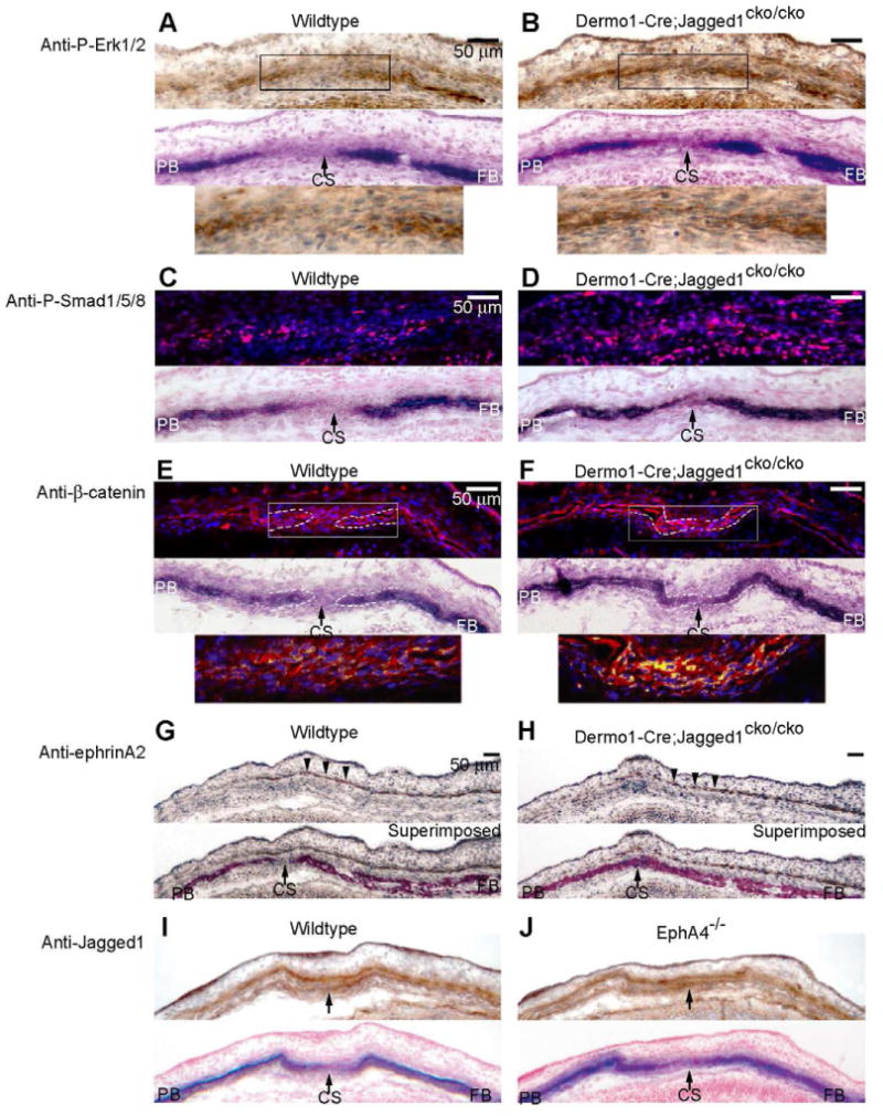
We examined the influence of mesoderm-specific inactivation of Jagged1 on the distribution of P-Erk1/2 (A, B), P-Smad1/5/8 (C, D), β-catenin (E, F) and ephrinA2 (G, H) in the coronal suture. (I, J) We also assessed the influence of loss of EphA4 function on Jagged1 expression. Wildtype and mutant embryos at E14.5 were prepared as in Fig. 1 and sections were stained with indicated antibodies. For P-Erk1/2, ephrinA2 and Jagged1, the antibodies were visualized with DAB (brown color) and sections were counterstained with hematoxylin. For P-Smad1/5/8 and β-catenin, the antibodies were visualized with Rhodamine Red-X-conjugated secondary antibody (red color). Nuclei were stained with DAPI (blue). In the case of β-catenin, the pink color of DAPI, β-catenin double positive nuclei was transformed to yellow to make it more visible. ALP stains of adjacent sections are shown below images of immunostains. The lower panels in A, B and E, F are enlargements of the boxed areas in the upper panels. Note that in wildtype embryos, P-Erk1/2, P-Smad1/5/8 were expressed in prospective bone and osteogenic fronts but not in the suture. β-catenin was expressed in the suture but was largely cytoplasmic. In Dermo1-Cre;Jagged1cko/cko mutants, P-Erk1/2 expression was coincident with ALP expression in the frontal and parietal bones. In contrast, P-Smad1/5/8 expression expanded into the sutural mesenchyme, similar to Notch2 and Hes1 expression (Fig. 3D, F), and ephrinA2 expression was unaltered (G, H). Jagged1 expression did not change in the EphA4-/- mutant coronal suture (I, J). CS, coronal suture; FB, frontal bone; PB, parietal bone. Scale bars: 50 μm.