
Figure 10.
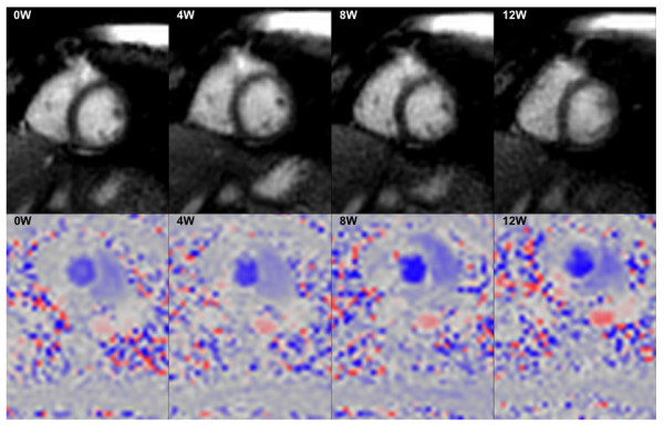
Real time data. Short-axis ventricular volumes (top) and flow data (bottom) acquired during increasing exercise within the MR scanner (0, 4, 8, 12W). Because the data is acquired in real-time, there is no need for the patient to attempt breath-holding during peak exercise, which is often difficult to achieve.