
Figure 3.
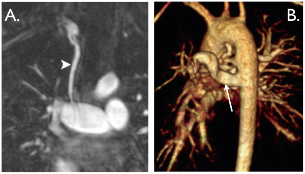
A. Coronal view from a contrast-enhanced MR angiogram showing a modified BT shunt (arrowhead). It originates from the innominate artery and inserts into a dilated right pulmonary artery. B. 3D, contrast-enhanced MR angiogram viewed from left posterior lateral showing several major aorto-pulmonary collateral arteries (MAPCAs). The arrow shows the largest MAPCA to the right lung.