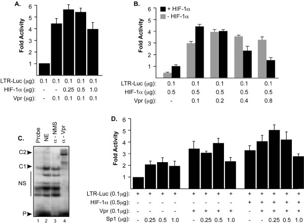
Figure 2.
Functional interplay between HIV-1 Vpr and HIF-1α. A and B. 786-O cells were transfected with LTR-Luc full length along with an increasing concentration of HIF-1α (A) and/or Vpr (B) expression plasmids. The amount of DNA in each transfection mixture was normalized with pcDNA6HisA. Luciferase activity was determined 48 hours after transfection. Results are displayed as histograms. C. Approximately 100, 000 cpm of synthetic [γ32P]-labeled double-stranded DNA oligonucleotide probe corresponding to the HIV-1 LTR GC-rich site was incubated with 10 μg of nuclear extracts prepared from microglial cells transfected with Vpr expression plasmid. Labeled probe was also incubated with nuclear extracts prepared from pcDNA-transfected microglial cells in the presence of anti-Vpr antibody (lane 4) and normal mouse serum (NMS) (lane 2). D. To further examine the interplay between Vpr and HIF-1α, 786-O cells were transfected with LTR-Luc along with HIF-1α, Vpr and/or Sp1 using different combinations as shown. Luciferase activity was determined 48 hours after transfection. Results are displayed as histogram. Each transfection was repeated 3 times using different DNA preps.

