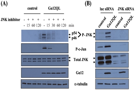
Figure 1. Gα12QL activates JNK in breast cancer cells.
MDA-MB-231 cells were infected with GFP (control) or Gα12QL adenovirus for 6–7 h. (A) Cells were starved for 2 h and then treated for the indicated time with JNK inhibitor SP600125 (20 µM) prior to lysis in JNK activation buffer and processing for analysis (see Materials and Methods ). (B) Cells were transfected with JNK siRNA or control (luc) siRNA for ∼72 h prior to infection with adenovirus as in (A). Cells were starved for 18 h before being lysed. For both panels, equal amounts of total protein in lysates were subjected to SDS-PAGE and immunoblot analysis to assess levels of phospho-JNK (P-JNK, p54 and p46) and phospho-c-Jun (P-c-Jun). Total levels of JNK and Gα12 expression, along with α-tubulin as a loading control, are also shown. Each panel shows the results of a single experiment that is representative of three or more independent experiments.

