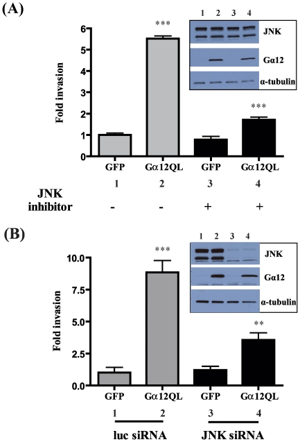
Figure 2. Gα12QL promotes cell invasion through activation of JNK.
MDA-MB-231 cells were infected with GFP (control) or Gα12QL adenovirus for 6–7 h and starved for ∼20 h. (A) Cells were pre-treated for 2 h with JNK inhibitor SP600125 (20 µM) and allowed to invade Matrigel for 24 h, in the presence of the inhibitor or vehicle control, in a transwell invasion assay (see Materials and Methods ). (B) Cells were transfected with JNK siRNA or control (luc) siRNA for ∼72 h before being infected with adenovirus. Cells expressing Gα12QL or GFP were allowed to invade Matrigel for 24 h. For both panels, the invasion assay was set up towards 5 µg/ml fibronectin. Cells that invaded were stained and counted in four random optical fields for each transwell, with three transwells per condition. Results are expressed as fold change in invasion compared to vehicle-treated GFP control cells. The insets show immunoblot analysis of the levels of Gα12, JNK and α-tubulin (loading control). Data is presented as mean ± SE from a single experiment that is representative of three independent experiments. ** p<0.01 and *** p<0.001, as determined by one-way ANOVA, followed by Tukey's multiple comparison test to obtain p values.

