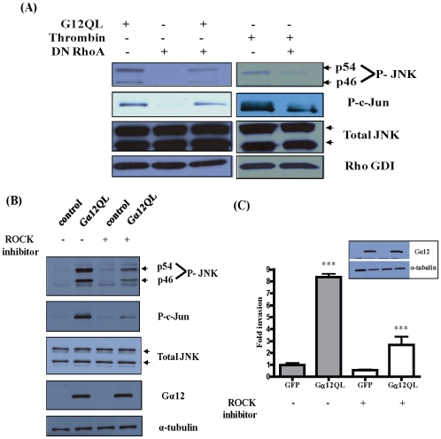
Figure 3. Gα12QL activates a ROCK-JNK signaling axis.
MDA-MB-231 cells were infected with adenovirus harboring GFP (control), Gα12QL or dominant-negative RhoA (DN RhoA) for 6–7 h. (A) Cells were starved and treated with 1 U/ml thrombin for 10 min, as indicated, before lysis in JNK activation buffer. Equal amounts of total protein in lysates were subjected to SDS-PAGE and immunoblot analysis to assess levels of phospho-JNK (P-JNK, p54 and p46) and phospho-c-Jun (P-c-Jun). Total level of JNK expression, along with Rho GDI as a loading control, is also shown. (B) Cells were starved and treated with ROCK inhibitor Y-27632 (10 µM) for 2 h before lysis in JNK activation buffer. Equal amounts of total protein in lysates were subjected to SDS-PAGE and immunoblot analysis to assess levels of phospho-JNK (P-JNK, p54 and p46) and phospho-c-Jun (P-c-Jun). Total levels of JNK and Gα12 expression, along with α-tubulin as a loading control, are also shown. Data shown in (A) and (B) are from a single experiment that is representative of three independent experiments. (C) Cells infected with GFP or Gα12QL adenovirus were starved for ∼18 h, then pre-treated with Y-27632 (10 µM) for 2 h and allowed to invade Matrigel for 24 h in the presence of the inhibitor, towards 5 µg/ml fibronectin. Cells that invaded were stained and counted in four random optical fields for each transwell, with three transwells per condition. Results are expressed as fold change in invasion compared to vehicle-treated GFP control cells. The inset shows immunoblot analysis of Gα12 and α-tubulin (loading control) levels. Data is presented as mean ± SE from a single experiment that is representative of three independent experiments. *** p<0.001, as determined by one-way ANOVA, followed by Tukey's multiple comparison test to obtain p values.

