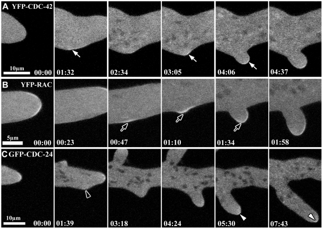
Figure 10. CDC-42 and RAC participate in lateral branch initiation.
Time series taken by laser scanning confocal microscopy of YFP-CDC-42, YFP-RAC and GFP-CDC-24 during lateral branching emergence. (A–B) Accumulation of CDC-42 and RAC at subapical region of the plasma membrane prior to the emergence of the new branch and after of establishment of a new axis of polarity are indicated by white and black arrows, respectively. (C) CDC-24 accumulation was not detected close to the plasma membrane (black arrowhead), but only as a cytoplasmic cloud, occupying the apical dome in the new branch as indicate the white arrowheads.

