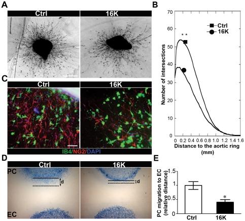
Figure 3. 16K hPRL inhibits pericyte outgrowth in an aortic ring assay and pericyte migration towards endothelial cells.
(A) Photomicrographs of mouse aortic ring cultured in collagen for 9 days and incubated without specific treatment (Ctrl) or under 16K hPRL treatment (16K). Data representative of at least 2 independent experiments are shown. Bar, 1 mm. (B) Quantification of migrating cell distribution around the aortic ring performed by computerized image analysis. Cell distribution is defined as the number of intersections between spreading cells and a grid of concentric rings, plotted as a function of the distance to the aortic fragment. Each curve is a mean of the cell network distribution obtained by averaging at least 5 individual distributions generated for each experimental condition. (C) Photomicrographs of a mouse aortic ring cultured in collagen for 9 days without specific treatment (Ctrl) or in the presence of 100 nM 16K hPRL. The rings were fixed for subsequent immunostaining: anti-Isolectin B4 Ab (IB4: green) identifying EC, anti-NG2 proteoglycan Ab (NG2: red) identifying PC/SMC. Nuclei were colored with DAPI (blue). Data representative of at least 2 independent experiments are shown. Bar, 100 µm. (D) HBVP migration towards HUVEC was assessed in an under-agarose migration coculture assay. HUVEC were treated for 72 h with 100 nM 16K hPRL (16K). d = migration distance between the beginning (solid line) and the end of the migration (dotted line). *, P<0.05.

