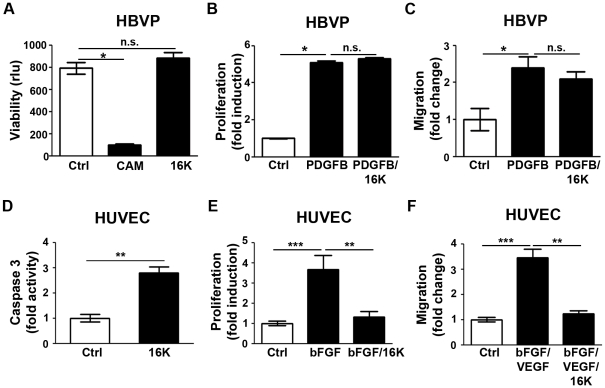
Figure 4. Endothelial cell specific effect of 16K hPRL.
The response of human brain vascular pericytes (HBVP) and human umbilical vein endothelial cells (HUVEC) to 16K hPRL was examined. (A) HBVP viability was evaluated by means of the calcein assay. 16K: 100 nM 16K hPRL. Cam: camptothecin-treated cells, a positive control. Rlu: relative luminescence units. Each bar represents a mean ± SEM, n = 3. Two different experiments were performed. (B) HBVP were treated with 20 ng/ml PDGF-BB to stimulate proliferation and 100 nM 16K hPRL (16K). HBVP proliferation was assayed 48 h later by measuring BrdU incorporation. The data presented are means ± SEM, n = 5 and are representative of at least two independent experiments. (C) HBVP migration was assessed in a modified Boyden chamber (Costar, Corning Inc.). HBVP were treated for 17 h with 20 ng/ml PDGF-BB to stimulate migration and 100 nM 16K hPRL (16K). The data presented are means ± SEM, n = 3 and are representative of at least two independent experiments. (D) Apoptosis in HUVEC was assessed by quantification of Caspase-3 activity. HUVEC were treated for 16h with 50 nM 16K hPRL. (E) HUVEC were treated with 10 ng/ml bFGF to stimulate proliferation and 100 nM 16K hPRL (16K). HUVEC proliferation was assayed 24 h later by measuring BrdU incorporation. The data presented are means ± SEM, n = 5 and are representative of at least two independent experiments. (F) HUVEC migration in a scratch-wound assay 8 h after treatment with bFGF and VEGFA and with or without 50 nM 16K hPRL. The data presented are means ± SEM, 5 measurements/condition, n = 3 and are representative of at least two independent experiments. n.s., non significant.*, P<0.05. **, P<0.001. ***, P<0.0001.

