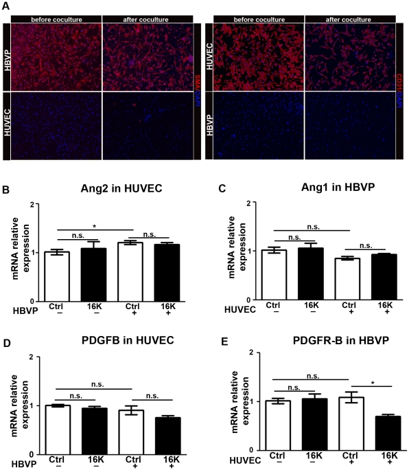
Figure 5. Decreased expression of PDGFR-B in HBVP cocultured with HUVEC after 16K hPRL treatment.
A. CD31 (endothelial cells) and α-SMA(pericytes) immunohistochemistry on HUVEC and HBVP cells before and after separation with MACS. (B-E) HUVEC pretreated for 30 min with 100 nM 16K hPRL (16K) were cultured for 16 h in the presence or absence of HBVP. The two cell populations were then separated prior to qRT-PCR analysis and subjected to qRT-PCR analysis to detect ANG2, ANG1, PDGFB and PDGFR-B transcripts. Data are presented as means ± SEM, n = 3 and are representative of at least two different experiments. n.s., non significant. *, P<0.05.

