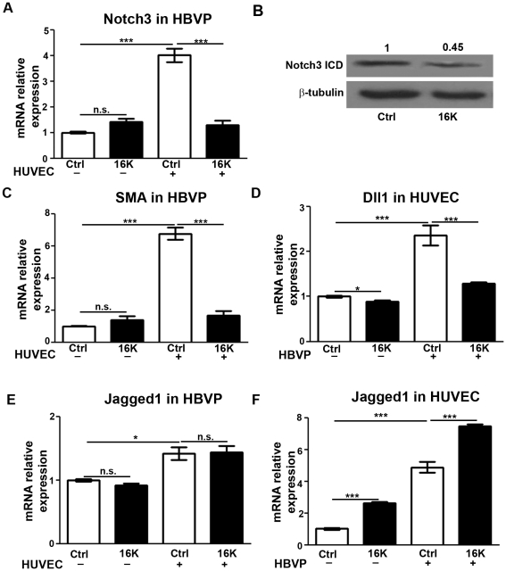
Figure 7. Decreased expression of Notch factors in HUVEC-HBVP cocultures after 16K hPRL treatment.
In cultures of HBVP or HUVEC alone, the cells were treated with 100 nM 16K hPRL (16K) for 16 h. HBVP+HUVEC cocultures were seeded and treated as in Fig. 6, but the time spent in coculture was longer (16 h). The two cell populations were then separated prior to qRT-PCR analysis. In HBVP, transcripts of NOTCH3 and aSMA were detected (respectively A and C). Western blot analysis was also performed to detect Notch3 ICD (B). In HUVEC, DLL1 transcripts were detected (D). In HBVP and HUVEC, JAGGED1 transcripts were detected (respectively E and F). The presented data are means ± SEM, n = 3 and are representative of at least two independent experiments. n.s., non significant. ***, P<0.0001.

