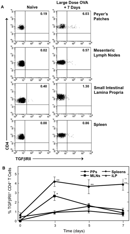
Figure 1. Effect of large-dose OVA feeding on the occurrence of CD4+ TGFβRII+ T cells.
Mononuclear cells were isolated from PPs, MLNs, iLP and spleens of naïve C57BL/6 mice (0) and at 24 h time points after 30 mg of OVA feeding. Cells were then stained with FITC-conjugated anti-hTGFβRII, APC-labeled anti-CD4, and biotin-tagged anti-CD3 mAbs followed by PerCP-Cy™5.5-conjugated streptavidin. Samples were subjected to flow cytometry analysis by FACSCalibur®. A. Plots are representative of naïve tissues and tissues analyzed 7 days after OVA feeding. B, Time course of CD4+ TGFβRII+ T cells in PPs, MLNs, iLP and spleens 3, 5 and 7 days after 30 mg of OVA feeding, n = 20 mice per time point, *p<0.05 or **p<0.001 when compared to sham-tolerized control group.

