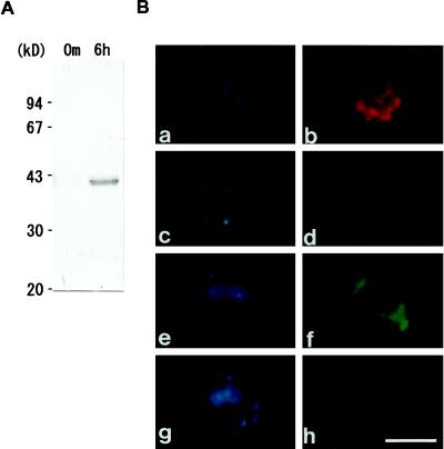
Figure 7.
A, Specificity of α-Zysfuse3, an antibody raised against affinity-purified, bacterially synthesized fusion protein of Zys3 and a 6× His tag. The fractionated total protein of the C. reinhardtii zygote at 0 min and 6 h after mating by SDS-PAGE was blotted onto the nitrocellulose membrane. The α-Zysfuse3 antibody was shown by western-blot analysis to specifically react with the 40-kD Zys3 proteins and the precursor in the zygote at 6 h. B, Indirect fluorescence microscopy for analysis of localization of Zys3 products. α-Zysfuse3 was used as the primary antibody in the mt− gamete (a and b), and in the zygotes at 1 h (c and d) and 6 h (e and f) after fertilization to detect its selective reaction against Zys3 proteins. Preimmune serum was also applied to zygotes at 6 h (g and h). Cells were observed under UV light by staining with DAPI (a, c, e, and g) and under blue light to detect the FITC signal (b, d, f, and h). Strong fluorescence of FITC was observed only around the cell nucleus from zygotes 6 h after fertilization. Bar = 5 μm. Red autofluorescence in b and d resulted from remnant chlorophyll molecules of the specimens.

