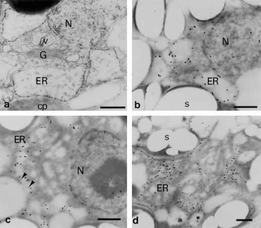
Figure 8.
Immunoelectron microscopic analyses with the anti-Zys3 protein antibody α-Zysfuse3. A general ultrastructural image of a very young zygote of C. reinhardtii is represented first as a control (a), and then at 3 h (b) and 6 h (c and d) after being reacted with α-Zysfuse3. N, Nucleus; cp, chloroplast; s, starch grain; G, Golgi apparatus; v, Golgi-related vesicle. Arrowheads indicate undefined structures protruding from the cytosolic side into the ER. Bars = 500 nm.

