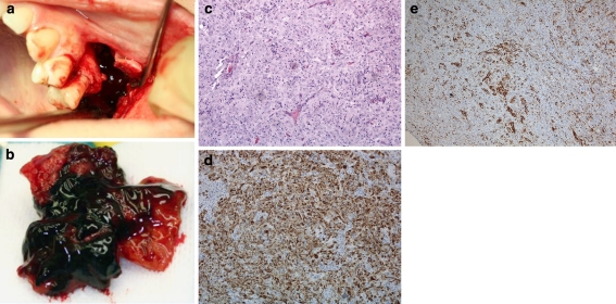
Fig. 2.
a Photograph of the left maxilla operation area of case 1. b Tumor mass from the left maxillary sinus. c The tumor is comprised of undifferentiated cells without connective tissue stroma. Varying degree of cellular and nuclear pleomorphism is noted on various sections. Intermixed are scattered small lymphoid cells (H&E, 40×). d Strong staining for cytokeratin verifies the epithelial origin of the tumour (AE1/AE3, 40×). e The lymphoplasmacytic infiltrate is highlighted by immunohistochemical staining with CD45 (20×)

