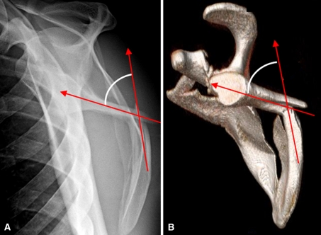
Fig. 3A–B.
Measurement of angulation on (A) a transcapular Y radiograph and (B) a 3D CT scan is demonstrated. A line is drawn through the proximal fragment in parallel with the cortices just proximal to the fracture. A second line is drawn through the distal fragment in parallel with the cortices just distal to the fracture. The angle formed by these two intersecting lines represents the angulation Note, even though the inferior teardrop forms a concave surface over the rib cage, it is the more proximal straight portion of the intramedullary canal that is used for the measurement (a critical distinction in measuring angular deformity so as not to overcall the angulation). 3D = three-dimensional.

