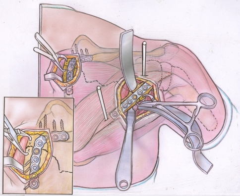
Fig. 2.
A diagram illustrates deep dissection and fixation. Lateral border dissection is through the interval between the teres minor and infraspinatus. The fracture along the medial border is exposed by localized elevation of the infraspinatus. Schanz pins are placed in the proximal and distal fragment (typically the neck and body) and used as joysticks to manipulate the fragments into the correct anatomic alignment. Pointed bone reduction forceps hold the fragments in the desired position until the plates are placed. The inset shows the screw vectors aimed into the area of the scapula with the best bone stock. Note the threaded screw hole overlying the fracture at the medial and lateral borders, indicating locking plates were employed for stable fixation.

