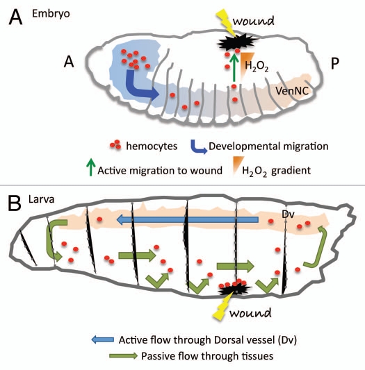
Figure 1.
Hemocytes migration during development and wound healing. (A) During the embryonic phase hemocytes (red dots) migrate following precise routes that distribute them in three parallel rows, following the ventral nerve cord (VenNC). At this stage the cue for their migration is represented by Pvr signaling (see text). In response to H2O2 production at the wounded site hemocytes divert from their conventional routes of migration to accumulate at the damaged tissue and initiate a wound healing response. A, anterior; P, posterior. (B) At the larval stage hemocytes are pumped in circulation by the dorsal vessel (Dv) that with its contractions ensures the hemolymph circulates with a posterior-to-anterior directionality (blue arrow). The opposite-oriented flow is rather a slower, passive one (green arrow). Hemocytes are thought to adhere to the damaged tissues once they randomly bump into them. The chemotactic signal at the wound at the larval stage has not been identified yet.

