
Figure 1. Organization of regions in the gastrointestinal tract.
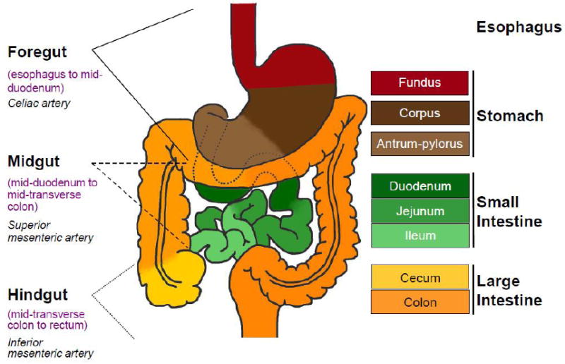
Left: The foregut, midgut and hindgut are classically defined according to the blood supply, as indicated. Right: Colors in the diagram refer to distinctive epithelia within the alimentary canal. In mice, the stratified squamous epithelium of the esophagus (crimson) extends into the dome-like forestomach, before forming a sharp, single-cell boundary with the glandular columnar epithelium of the gastric corpus (dark brown). As discussed in the text, the boundary between the gastric body and antral-pyloric epithelia (light brown) is less distinct, while that between the stomach and small intestine (green), marked by the pyloric sphincter, is sharp. Small intestine regions have different digestive functions and characteristic patterns of genes expression, without well-defined boundaries. The villous epithelium of small intestine transitions abruptly into a flat, non-villous epithelium at the ileo-cecal valve, which is followed by a specialized cecum (yellow) and the remainder of the colon (orange).