
Figure 3. Illustrative boundary defects in mutant mice.
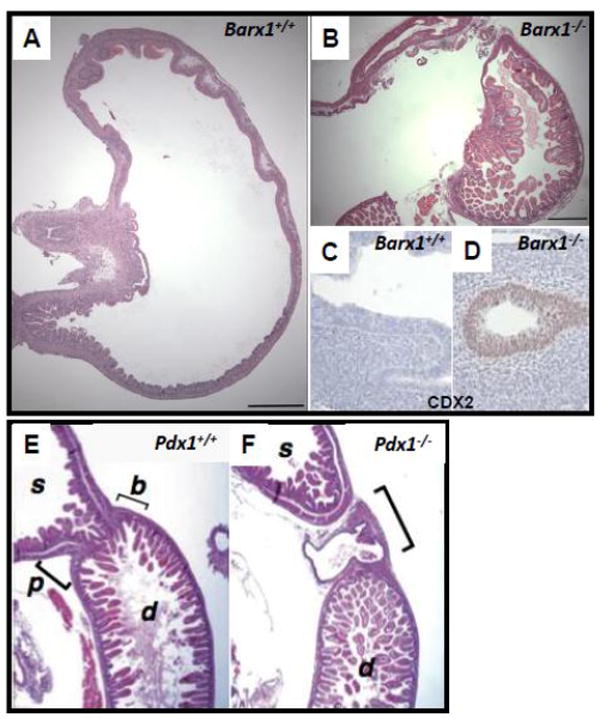
(A-D) Hematoxylin and eosin (H&E)-stained neonatal stomach tissues from wild-type (A) and Barx1-/- (B) mice [52]. The homeotic posteriorization in Barx1-/- stomach is evident from ectopic presence of intestinal villi (B) and expression of the intestinal marker CDX2 (D) as early as E12.5 [27]. (E, F) H&E-stained wild-type (E) and Pdx1-/- (F) tissues at E18.5 reveal defective pylorus development in Pdx1-/- mice. Normally the stomach (s) opens into the duodenum (d) at the pylorus (p), where well-defined Brunner’s glands (b) are found. In Pdx1-/- mutants, a cavity that lacks villi and is lined by a cuboidal epithelium forms in this region [23].