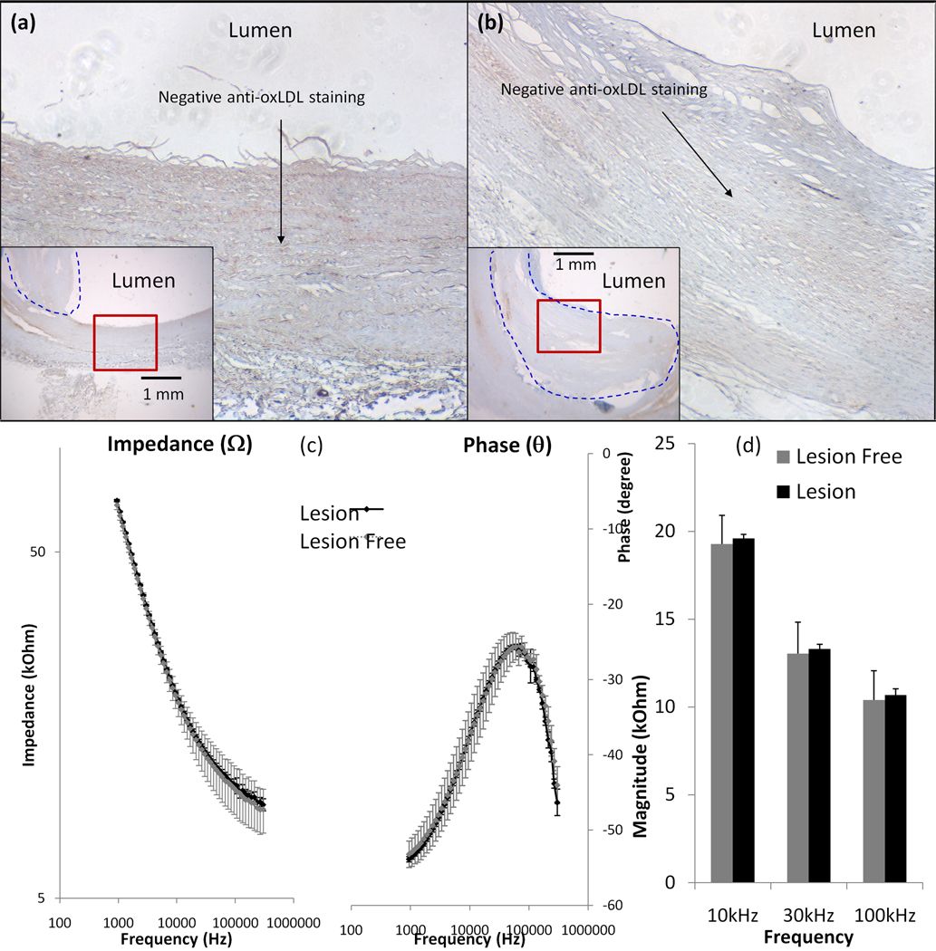
Figure 4. Endoluminal EIS measurements of oxLDL-absent fibrous atheroma.
(a) Anti-oxLDL staining was negative in the lesion-free site. (b) Anti-oxLDL staining was also negative in the fibrous atheroma. (c) Frequency-dependent endoluminal tissue impedance from 1 kHz to 300 kHz between lesion-free regions and fibrous structures were statistically insignificant. (d) Bar graph also revealed insignificant difference in frequency-dependent impedance from 10 kHz to 100 kHz (p > 0.05, n = 4 for lesion-free and n = 6 for oxLDL-absent fibrous atheroma).

