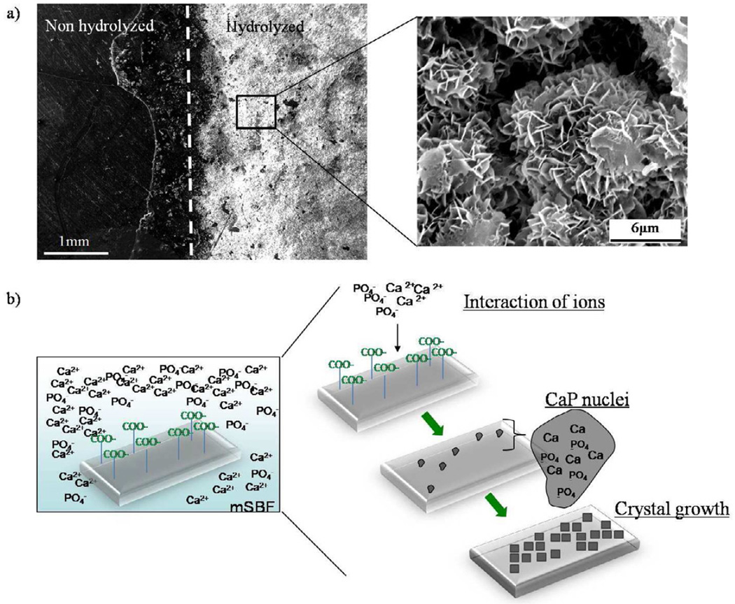
Figure 4.
a)Hydrolysis of PCL. Partial exposure of PCL films to NaOH resulted in the formation of a mineral coating in the region exposed to the hydrolysis pre-treatment. The surface that was protected from the NaOH solution did not show evidence of mineral formation after incubation for 7 days in mSBFlow HCO3. b) Schematic representation of the process of mineral coating formation after incubation in simulated body fluids. Hydrolysis of PCL scaffolds prior mSBF incubation results in an increase in the number of carboxylic acid groups on the surface of the scaffold. These negatively charged groups interact with calcium and phosphate ions in solution to form CaP rich nuclei from which the mineral grows creating a mineral coated surface.

