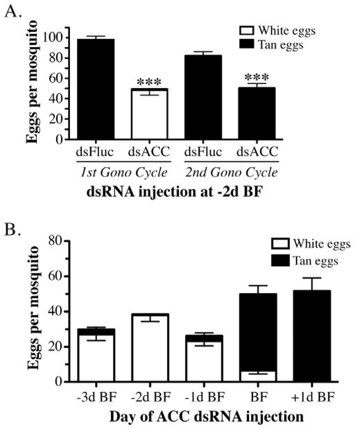
Figure 6.
Quantitation of egg production in ACC deficient mosquitoes. A) Data collected from mosquitoes after the 1st and 2nd gonotrophic cycle that had been injected with 1.0 μg of dsFluc or dsACC RNA at 2 days prior to the first blood feeding. Each bar represents the total number of eggs laid per mosquito (Mean ± SEM), which were scored at 4 hrs post-oviposition as inviable white eggs (white fill) or viable tan eggs (black fill). Asterisks indicate a significant difference between the total number of eggs oviposited per ACC deficient mosquito compared to the control Fluc dsRNA injected mosquitoes (***P <0.001). B) Number of white and tan eggs oviposited per dsACC RNA injected mosquito at various times before and after the blood meal plotted as Mean ± SEM. Data in A and B were derived from three independent biological replicas.

