Figure 9.
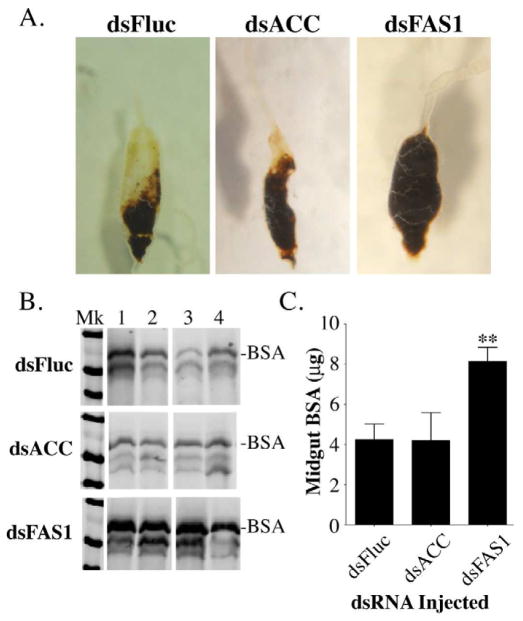
Blood meal digestion is delayed in FAS1 deficient mosquitoes. A) Representative photos of midgut tissues from dsFluc, dsACC, and dsFAS1 RNA injected mosquitoes dissected 48 hours after blood feeding. B) Representative western blot of midgut extracts prepared from four individual dsFluc, dsACC, and dsFAS1 RNA injected mosquitoes showing the extent of BSA degradation at 48 hr PBM. C) Quantitative analysis of BSA degradation data using pooled mosquito midguts isolated at 48 hr PBM from dsFluc, dsACC, and dsFAS1 RNA injected mosquitoes. Data were collected from three independent biological replicas using image analysis of BSA western blots and plotted as Mean ± SEM as previously described (Isoe et al., 2009). Asterisks indicate a significant difference between the amount of BSA remaining in midguts from dsFAS1 and dsFluc RNA injected mosquitoes (**P <0.01). In contrast, there was no significant difference between the amount of BSA remaining in midguts from dsACC and dsFluc RNA injected mosquitoes.

