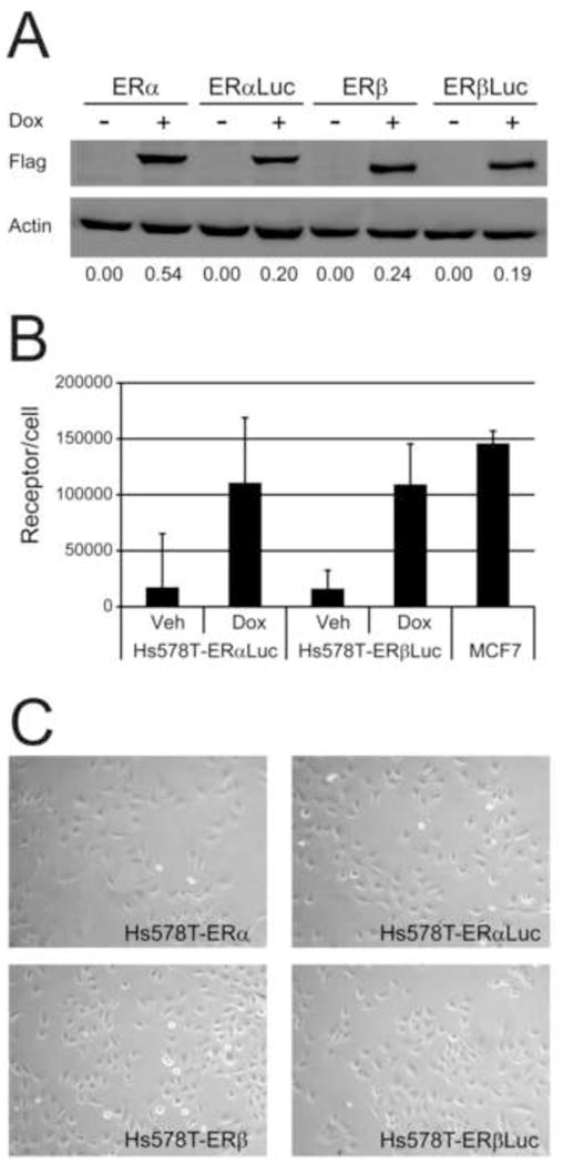
Figure 3.

Hs578T-ERαLuc and Hs578T-ERβLuc cells express similar levels of ER. (A) Quantitative western blot with Hs578T-ERα (ERα), Hs578T-ERαLuc (ERαLuc), Hs578T-ERβ (ERβ), and Hs578T-ERβLuc (ERβLuc) treated with vehicle (-Dox) or 50 ng/mL Dox (+Dox). ER expression was detected using FLAG antibody and quantified by normalizing to b-actin using the Licor Odyssey near-infrared gel reader. The normalized integrated intensity for the FLAG signal is shown below the images. (B) Ligand binding assays confirmed the quantitative western blots. Hs578T-ERαLuc and Hs578T-ERβLuc cells were seeded in triplicate and treated with vehicle or 50 ng/mL Dox for 24 hr. Cells were labeled with 20 nM [3H]-E2 in the presence or absence of cold competitor for 2 hr, washed, and total cell lysate was assessed for bound radioactivity as described in Materials and Methods. MCF7 cells were included for comparison. Two additional wells of each cell line and condition were used to determine the cell number and the numbers of receptors per cell were calculated based on a 1:1 molar ratio of ligand to receptor. The average and standard deviation of three independent experiments are shown. (C) The morphology of Hs578T-ERαLuc and Hs578T-ERβLuc was similar to that of the parent Hs578T-ERα and Hs578T-ERβ cell lines. Representative phase-contrast microscopy images of each cell line (100X magnification).
