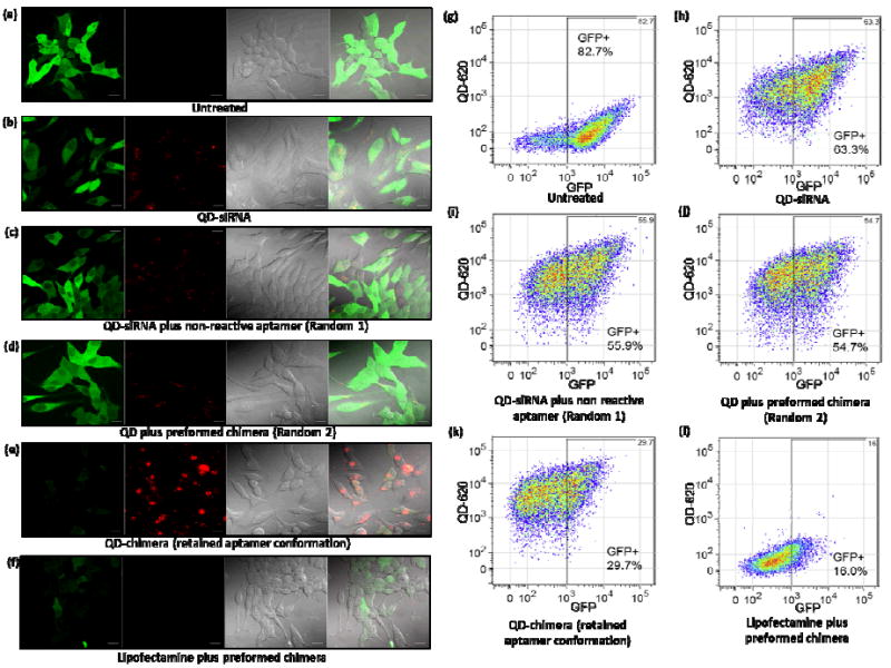
Figure 4.

Evaluation of eGFP gene silencing in C4-2B cells with confocal microscopy and flow cytometry. (a) Untreated C4-2B cells only, (b) cells treated with QD-siRNA without aptamer, (c) cells treated with QD-siRNA and aptamer without terminal thiol group (random 1), (d) cells treated with QD and preformed chimera (random 2), (e) cells treated with QD-chimera with retained aptamer conformation, and (f) cells treated with Lipofectamine-chimera. Fluorescence imaging shows significant GFP reduction (green) and QD fluorescence (red) in (e). (g-k) Quantitative flow cytometry of eGFP silencing corresponding to the above fluorescence imaging studies. At the current gate value, the eGFP-positive cells are 82.7%, 63.3%, 55.9%, 54.7%, and 29.7%, respectively, revealing enhanced silencing is only observed when aptamer is in its native conformation. The flow cytometry study also confirms the microscopy result on enhanced uptake of QD-chimera with intact conformation. Compared to cells treated with QD-siRNA without targeting aptamer (h), cells in random 1 (i), random 2 (j), and QD-chimera (k) experiments on average uptake approximately 1.4, 1.4, and 2 times more particles revealed by the QD fluorescence channel. (l) Positive control using Lipofectamine confirms the activity of the chimera.
