
Figure 5.
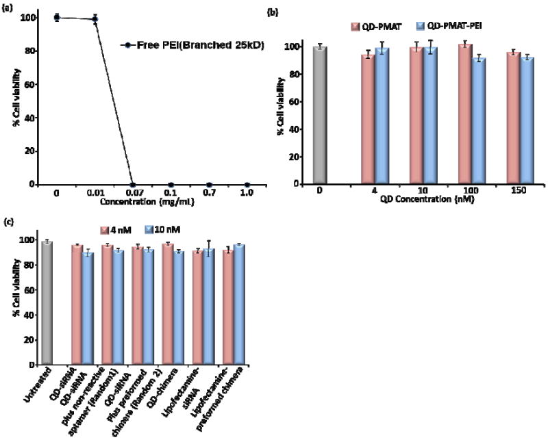
Dose-dependent cytoxicity of free PEI and QD-PMAT-PEI nanoparticles in C4-2B cells after 24 h treatment. (a) free PEI shows a sharp decline in cell viability above 0.4 μM. (b) QD-PMAT and QD-PMAT-PEI nanoparticles are not toxic to cells upto 150 nM based on QDs (for QD-PMAT-PEI the PEI concentration is 1.2 μM). (c) Comparison of cytotoxicity of QD-PMAT-PEI (4 and 10 nM) in the presence of siRNA. Cell viabilities after treatment with QD-siRNA, random 1, random 2, QD-chimera with preserved conformation, Lipofectamine with siRNA and Lipofectamine with preformed chimera, compared with untreated cells. The assembled complexes at the above concentrations are non-toxic to cells.