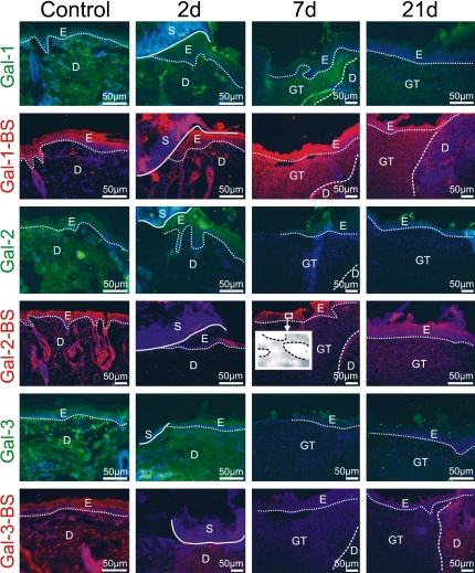
Fig. 2.
Illustration of immunohistochemical galectin detection and localization of accessible binding sites (BS) for labeled galectins in the epidermis and in the dermis/granulation tissue during healing. Comparison between control data (first vertical panel) and specimens at each studied time point during healing (2d: second vertical panel; 7d: third vertical panel; 21d: fourth vertical panel) is thus made possible for each marker along each horizontal panel. In detail, the following assignments of type of probe and time point are given. First panel (Gal-1): strong signal intensity for galectin-1 two days after injury in both epidermis and dermis near the wound edge, decreasing over time to minimal presence in the dermis at day 21; second panel (Gal-1-BS): low level of galectin-1 reactivity in uninjured skin and wounds two and 21 days post wounding, increased reactivity to Gal-1-BS in the granulation tissue; third panel (Gal-2): galectin-2 detection in the epidermis, absence in granulation tissue; fourth panel (Gal-2-BS): galectin-2 reactivity in uninjured skin and wounds localized to epidermis, low-level presence of binding sites in the dermis, insert—galectin-2 nuclear reactivity in the epidermis (dashed line marks the nuclei of keratinocytes); fifth panel (Gal-3): presence in the suprabasal epidermal layer and dermis, low-level signal intensity in the granulation tissue; sixth panel Gal-3-BS: present in the suprabasal epidermal layer and in the surrounding dermis, low abundance presence in the scar forming. For orientation solid/dotted/broken lines are given separating distinct regions referred by the following abbreviations: E, epidermis; D, dermis; GT, granulation tissue; S, scab; in detail, a dotted line sets epidermis apart from dermis and/or granulation tissue; the broken line distinguishes dermis from granulation tissue and the solid line scab/necrosis from vital tissue.

