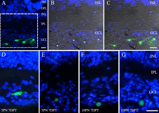
Fig. 4.
Integration of transplanted bone marrow-derived mesenchymal stem cells. (A) GFP-expressing cells integrated throughout the 5PN 7DPT host retina. (B, C) Higher magnification view of the boxed area in (A). Merged image of differential interference contrast micrograph, DAPI, and GFP staining pictures of the bone marrow-derived mesenchymal stem cells in ganglion cell layer. Asterisks indicate GFP-positive cell bodies. (D, E) The transplanted cells integrated into the ganglion cell layer at 5PN 7DPT retina. (F, G) The transplanted cell integrated into the ganglion cell layer at 10PN 7DPT retina. Bar=20 µm.

