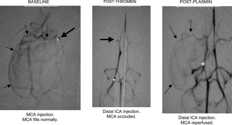
Figure 7.
Middle cerebral artery (MCA) occlusion in the rabbit model of ischemic stroke.12 The baseline study (left panel) shows the tip of the microcatheter (heavy arrow) and course of the MCA (light arrows). After local infusion of thrombin, only the M1 segment of the MCA is filled by contrast (middle panel, heavy arrow). Following catheter delivery of plasmin (2 mg) to the distal ICA, the MCA is reperfused (light arrows, right panel). Reproduced with permission from Jahan R, Stewart D, Vinters HV, et al. Middle cerebral artery occlusion in the rabbit using selective angiography: application for assessment of thrombolysis. Stroke, in press. Lippincott, Williams & Wilkins.

