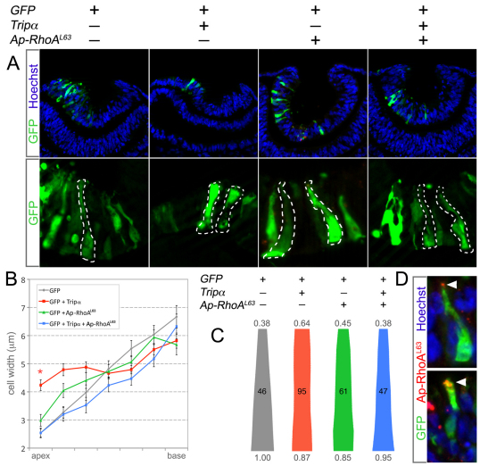
Fig. 5.
Trio activation of RhoA is required for apical constriction during lens pit invagination. (A) Stage 11 chicken embryos were electroporated in ovo with the indicated plasmid(s) and allowed to develop until the lens pits began to invaginate. Transgenic embryos were cryosectioned and the outline of GFP-positive cells was determined (dashed lines). (B) The average apical width along the apical basal axis of transgenic cells from each experimental group is depicted. Error bars represent s.e.m. and the asterisk identifies significant differences from the control (P<0.05). (C) The average cell shape from each experimental group is depicted. The number value above or below each shape is the fold difference between the average basal width of control cells and the apical or basal widths, respectively, of the transgenic cells. The number of cells analyzed for each group is indicated in the middle of each outline. (D) Electroporated chicken lens pit cells labeled for GFP (green), nuclei (blue) and epitope tagged Ap-RhoAL63 (red). The arrowheads indicate the apical localization of Ap-RhoAL63.

