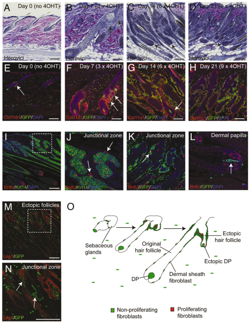
Fig. 7.
Remodelling of adult dermis originates from a population of fibroblasts in the hair follicle junctional zone. (A-L) Sections of K14β-catER mouse back skin treated with 4-OHT for 0, 7, 14 or 21 days as indicated or for 14 days (I-L). (A-D) Paraffin sections stained using Herovici’s method to identify mature (pink fibres) and immature (fine blue fibrils) collagen. (E-H) Frozen sections labelled with anti-Col11α1 (red) and showing GFP fluorescence (green). Nuclei were stained with DAPI (blue). (I-L) Paraffin sections stained for Krt14 (green) and BrdU (red). (M,N) Frozen section of ectopic follicle-forming K14β-catER back skin labelled with anti-Lrig1 (red) and showing GFP fluorescence (green). The boxed regions in I and M are enlarged in J and N, respectively. Arrows point to: (E) a small population of PdgfraEGFP+ fibroblasts associated with the telogen junctional zone; (B,F) regions of recently regenerated collagen at the junctional zone; (C,D,G,N) ectopic follicles; (J,K) BrdU-labelled junctional zone fibroblasts; and (L) the DP of an original hair follicle, which is not labelled with BrdU. (O) The progressive remodelling of adult dermis originating from fibroblasts associated with the hair follicle junctional zone. Scale bars: 200 μm.

