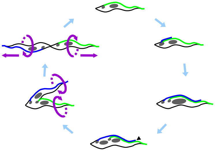
Fig. 7.
Cartoon depicting major stages of the Trypanosoma brucei cell division cycle in procyclic lifecycle stages. The cell cycle begins with replication of the basal body and initiation of new flagellum (blue) biogenesis. The new flagellum extends along a path defined by the old flagellum (green), while the basal bodies and associated kinetoplasts (small gray circles) migrate away from each other. In a closed mitotic cycle, the nucleus (large gray circle) is replicated and the new nucleus assumes a position between the new and old kinetoplast/basal body apparatus. Cytokinesis initiates at the tip of the new flagellum (black arrowhead) and cleavage furrow ingression proceeds to the cell posterior, separating the two daughter cells between the new and old flagellum. Ultimately, daughter cells are oriented in opposite directions, with their flagella exerting rotational and pulling forces that facilitate final cell separation.

