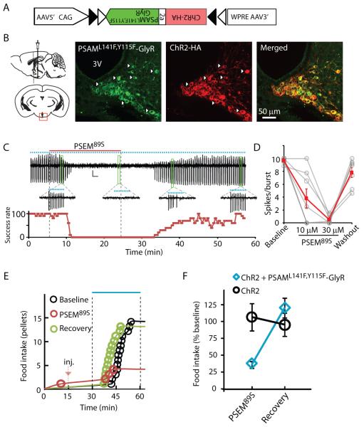
Fig. 4.
Stringent neuronal silencing test of PSAML141F,Y115F-GlyR for suppressing AGRP neuron-evoked feeding behavior. (A) Construct for a Cre-dependent recombinant adeno-associated viral vector with an inverted bicistronic open reading frame for PSAML141F,Y115F-GlyR and ChR2 under control of a FLEX-switch [two antiparallel pairs of heterotypic loxP sites (triangles)]. (B) Diagram illustrating injection site (left) and confocal images (right) taken from brain slices of virally transduced Agrp-cre mice showing co-expression of PSAML141F,Y115F-GlyR (green, α-Bgt-Alexa488) and ChR2 (red, anti-HA). (C) Cell-attached recording showing suppression of light-evoked bursts of action potential currents (10 Hz light pulses for 1 s, represented by blue dots) following application and then wash-out of PSEM89S (above). Bursts are expanded for selected time points. Below is spike success rate (percent) before, during and after PSEM89S application. (D) Successful light-evoked spikes per burst (n = 8 cells). (E) Food intake on successive days resulting from photostimulation in an Agrp-cre mouse co-expressing ChR2 and PSAML141F,Y115F-GlyR after saline (Baseline and Recovery) or PSEM89S [intraperitoneal injection (inj.), marked with red arrow]. (F) Evoked food intake normalized to initial photostimulation session. Injection of PSEM89S reduced photostimulation-evoked eating in Agrp-cre mice co-expressing ChR2 and PSAML141F,Y115F-GlyR (30 mg/kg, blue diamond, n = 5 mice) but not in Agrp-cre mice expressing only ChR2 (50 mg/kg, black circle, n = 6 mice). Error bars are s.e.m.

