
Figure 1.
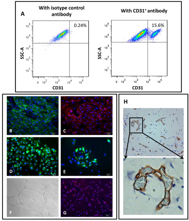
Derivation and characterization of hiPSC-ECs. A) [Left panel] Isotype control. [Right panel] FACS plot of data obtained using CD31+ antibody to quantitate ECs derived from hiPSCs B) to E) Immunofluorescent staining of the hiPSC-ECs for endothelial markers CD31, CD144, eNOS and vWF. F) hiPSC-ECs form a capillary-like network on Matrigel 24 hours after seeding the cells. G) hiPSC-ECs take up acetylated LDL. Scale bar: 50μm. H) in vivo formation of capillary structures in matrigel plugs after subcutaneous transplantation in SCID mice, stained using an Ab specific for human CD31.